(a) Bright field TEM image visualizing size and CeO2 nanoparticles;
(b) high resolution TEM image of structural modulations in Li-ion conductor Li3xNd2/3−xTiO3;
(c) HAADF-STEM image of the SnO2 whisker sensibilized with Pd nanoparticles;
(d) HAADF-STEM image of the carbon-coated SnO2 nanowire
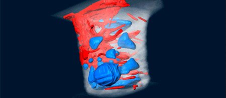
Bundle of carbon nanotubes (in red) grown from catalytic nanoparticles inside a Si contact hole.
The three-dimensional (3D) distribution of carbon nanotubes (CNTs) grown inside semiconductor contact holes is studied by electron tomography. The use of a specialized tomography holder results in an angular tilt range of +/-90 degrees which means that the so-called "missing wedge" is absent. The transmission electron microscopy (TEM) sample for this purpose consists of a micropillar that is prepared by a dedicated procedure using the focused ion beam (FIB) but keeping the CNTs intact.
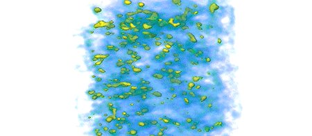
It is not straightforward to determine the exact location of nanoparticles in a 3D mesoporous network based on only 2D projections as is done in conventional TEM. Therefore, electron tomography was performed. This is a method in which a 3D structure is reconstructed from a series of (S)TEMimages acquired at successive tilts. It is confirmed that the anatase particles are located inside the porous structure of MCF.
Electron tomography was performed using a Fischione onaxis rotation tomography holder (model 2050). This holder is designed for nanopillar shaped samples, which were prepared using a DualBeam SEM/FIB system. The spatial resolution of the tomography experiment in this study was determined to be 0.6 nm. 3D reconstruction was carried out using conventional reconstruction algorithms as well as more advanced algorithms (total variation minimization) that facilitate the quantification of 3D data.