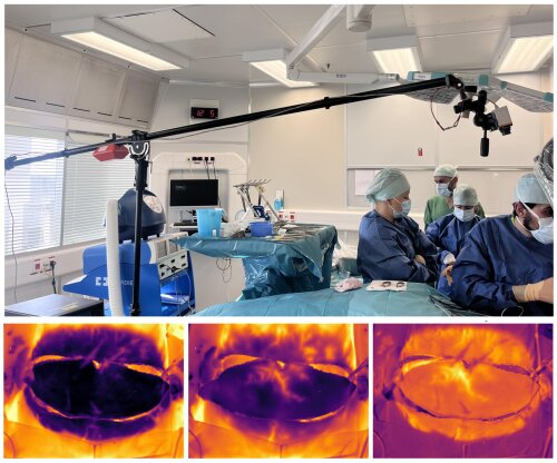
Well-executed breast reconstructions are hugely important for the mental well-being of cancer patients. In recent years, science has made a lot of progress with breast reconstruction using own tissue. Within this research, the focus is on minimising damage to the tissue origin site to avoid tissue death.
DIEP flaps
Today, breast reconstructions with so-called DIEP flaps (Deep Inferior Epigastric Artery Perforator) are preferred. This method causes less damage and produces beautiful aesthetic results. Choosing the right blood vessels for the flap is therefore essential because it is irrigated by only one perforator (= blood vessel starting from the muscles and reaching the surface). But the success of breast reconstruction does not only depend on the selection of the right blood vessels. Sometimes the flap fails due to damage during perforator dissection, failure of blood vessel coupling, pinching off or compression of the vessel stem during flap shaping. To diagnose these problems, we often use clinical monitoring.
Thermal imaging cameras
Research group InViLab at the University of Antwerp has been working for years with the University Hospital Antwerp (UZA) to evaluate thermal imaging cameras as a non-invasive technique when applied to all phases of breast reconstruction. This technique allows the surgeon to identify the most dominant perforators and determine the well-perforated area. Clinical studies already confirmed DIRT's ability to visualise the location of perforators of DIEP flaps and thereby minimise tissue mortality. Moreover, the use of DIRT (Dynamic InfraRed Thermography) provides additional information on perforator quality through objective monitoring of flap perfusion.
With your support, we can further intensify research so that surgery in cancer patients is optimal and we can avoid tissue mortality.