DAY 1 - Thu 7 May 2026
Royal Academy of Fine Arts Antwerp, Mutsaardstraat 31, Antwerp
Roberta Ballestriero (Italy)
From Flesh to Form – From Studio to Specimen: Artistic Anatomy and Scientific Illustration at the Academy of Fine Arts in Venice
Throughout the centuries, artists and scientists have been fascinated by the human body and the natural world, seeking to represent it in two and three dimensions. The need to create scientific and didactic illustrations—to understand and disseminate knowledge—has therefore fostered a union between art and science that has developed over time.
A particular drawing method, taught for several years at the Academy of Fine Arts in Venice (Accademia di Belle Arti di Venezia), is “Struttura Uomo”, which is strictly inductive and analytical. The approach to reproducing the human body is completely reversed, inverting the representative method by proceeding from inside to outside.
The intention of this course is to expand the student’s ability to see, understand, and represent the human body in its spatial, volumetric, and sculptural dimensions. It emphasizes that the role of an Academy of Fine Arts is not to teach how to copy forms, but to train the gaze—to “see inside the form” and “see-through”—as taught by the Venetian school in the “Visione Trasparente” (“Transparent View”).
Within the tradition of fine arts academies, copying and direct observation of original works have always been essential components of academic training. This practice is even more significant in the field of Scientific Illustration, where direct observation of natural specimens and living models—often preserved in scientific museums and botanical gardens—represents an irreplaceable element of learning.
Since 2022, thanks to the collaboration and availability of museum staff, students enrolled in the Scientific Illustration course have had the opportunity to visit the scientific museums of the University of Padua annually. This collaboration has gone beyond traditional guided visits, giving rise to specialized workshops and study days dedicated to drawing from life. This relationship, still in its early stages, was further consolidated in 2025 through a formal agreement between the University Museum Centre (Centro di Ateneo per i Musei, CAM) and the Academy of Fine Arts in Venice, aiming to enhance a shared educational heritage that enables students to become more aware, creative, and capable of communicating science through art.
Biography
Roberta Ballestriero obtained her European PhD from the Complutense University of Madrid. Since 2004, she has lectured in Art History at several British universities and currently serves as Art Historian in residence at the Gordon Museum of Pathology, London. She teaches at the Academy of Fine Arts in Venice and collaborates with the University of the Arts, London.
She is a Scientific Member of the Forensic Anthropology, Paleopathology and Bioarchaeology (FAPAB) Research Centre in Avola, Syracuse, and an Honorary Member of the Bologna Surgical Medical Society.
Her research explores the intersection of art and science, with a particular focus on the art of ceroplastics/wax modelling over the centuries. This includes anatomical models, portraits, ex-votos, wax sketches, and contemporary art. She founded and presided over the first international congresses on wax modelling in forty years (London 2017, Padua 2019, Mexico City 2025) and has edited the volumes Ceroplastics, The Art of Wax (2019) and Ceroplastics, The Science of Wax (2021).
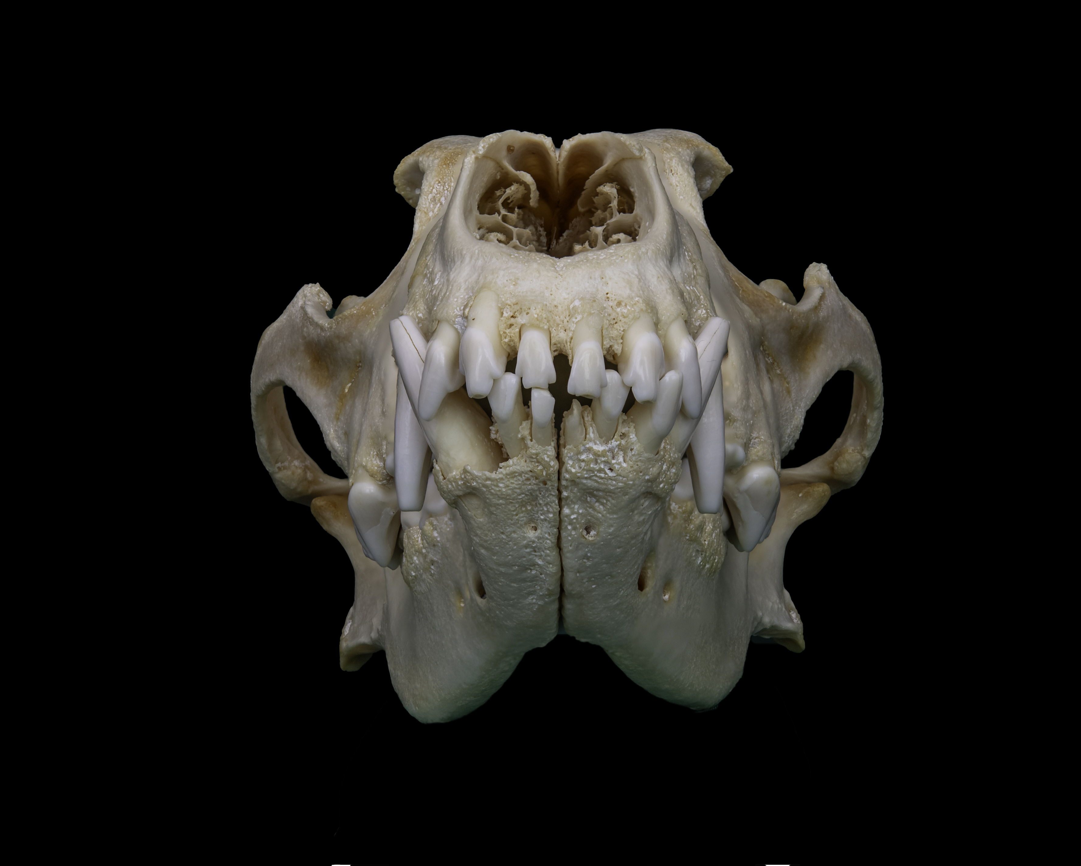
Figure. Skull of a German Shepherd dog (Canis lupus familiaris), Osteological collection of the Museum of Veterinary Medicine, University of Padua. Department of Comparative Biomedicine and Nutrition. Photo: R. Ballestriero, O. A. Burke.
Wendy Birch (UK)
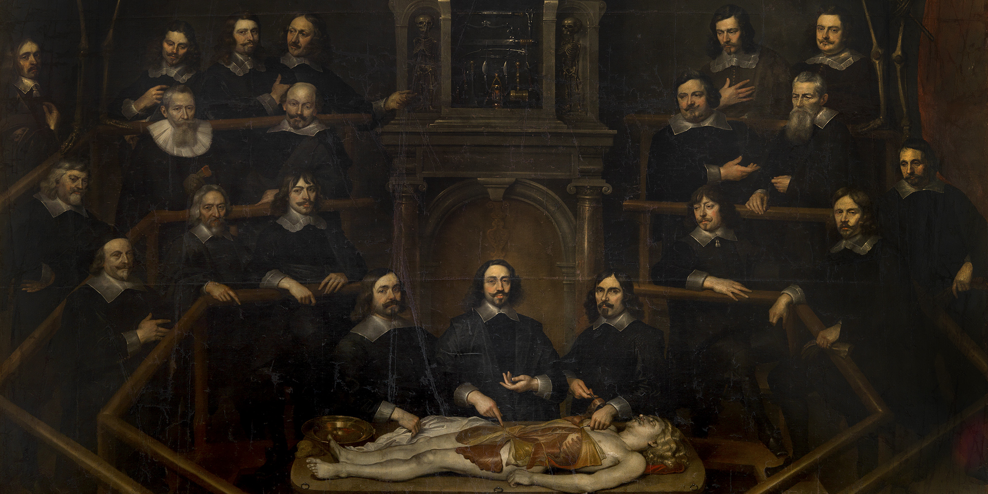
Dr. Tulp’s Lesson and the Dissection of the Forearm: From Painting to Practice
The Anatomy Lesson of Dr. Nicolaes Tulp by Rembrandt van Rijn remains one of the most compelling visual documents of anatomical teaching in early modern Europe. The painting depicts Nicolaes Tulp demonstrating the musculature of the left forearm during a public dissection of the executed criminal Aris Kindt, in the presence of members of the Amsterdam Guild of Surgeons. While widely admired for its dramatic composition and realism, the anatomical accuracy of the forearm has long been the subject of debate among both medical historians and art historians.
This lecture revisits these longstanding discussions by directly comparing the anatomy represented in the painting with findings from cadaveric forearm dissections. Moving beyond earlier interpretations that relied primarily on anatomical atlases, it focuses on several features often identified as problematic: the oblique muscular structure on the proximal ulnar forearm, the muscles held in Tulp’s forceps, the elongated muscle along the ulnar border, and the slender cord-like structure extending toward the little finger.
Rather than interpreting these elements as straightforward inaccuracies, this reassessment highlights the complex interplay between dissection practice, didactic demonstration, and artistic interpretation. The depicted structures correspond most closely to the flexor carpi radialis, flexor digitorum superficialis, and flexor carpi ulnaris muscles. The narrow cord-like structure along the ulnar side of the hand—often interpreted as a variant of the ulnar nerve—may alternatively represent the tendon of an accessory abductor digiti minimi muscle. This anatomical variant, whose tendon can traverse the hypothenar region toward the proximal phalanx of the little finger, aligns closely with the trajectory shown in the painting and offers a plausible explanation for the otherwise enigmatic “white cord,” though artistic stylisation cannot be excluded.
The lecture is followed by a practical session led by Francis Van Glabbeek and Tom Quisenaerts. In this hands-on component, participants will study dissected forearms to identify and critically assess the anatomical structures represented in Rembrandt’s work, creating a direct dialogue between historical imagery and contemporary anatomical observation.
Biography
Wendy Birch is a forensic anatomist and Associate Professor (Teaching) in Anatomy at University College London (UCL), where she also manages the UCL Anatomy Laboratory. She serves as the Designated Individual responsible for UCL’s Human Tissue Act anatomy licences and leads anatomy education within the UCL MBBS (Medicinae Baccalaureus, Baccalaureus Chirurgiae) programme.
Her academic interests span anatomical and forensic sciences, medical education, and the history and visual culture of anatomy. Her research focuses on how anatomical knowledge is communicated through teaching, art, and material culture, with particular attention to historical representations of dissection and the dynamic relationship between anatomical practice, visual representation, and medical education.
Figure: The Anatomy Lesson of Dr. Nicolaes Tulp, Rembrandt van Rijn, 1632, oil on canvas, Mauritshuis, The Hague, Netherlands.
Joanna Cameron (UK)
Medical Art – Observation and Techniques for the Medical Artist
Medical artistry has long been a source of inspiration and wonder. Core skills—rooted in careful observation and strong drawing ability—remain as essential today as in the past. This practice involves studying form, skeletal structures, and anatomy, while refining traditional art techniques, all with the goal of producing artwork with specific medical or educational purposes. By combining scientific understanding with artistic skill, medical artists translate complex anatomical knowledge into visually engaging and accurate representations that communicate, educate, and inspire.
Biography
Joanna Cameron is an artist and author with a lifelong passion for medical art, specializing in drawing, pen and ink, watercolour, and printmaking. She is the author of Medical Art – Principles and Techniques for the Creative Artist (2025).
Shortly after graduating in Medical Art from St. Bartholomew’s Hospital in 1988, she was invited to serve as Honorary Secretary of the Medical Artists’ Association Education Committee. She later supported the Medical Artists’ Education Trust as Director of Education from 2006 to 2022 and was awarded an MAA Fellowship in 2013 for her longstanding contributions to the MAET Charitable Trust. She now teaches the MAET Advanced Foundation Programme, focusing on drawing and observation, and became Chair of the Medical Artists’ Association of Great Britain (MAA) in 2025.
Beyond medical art, Joanna is passionate about the natural world. She wrote and illustrated the Wildlife and Nature Notes and the Heritage series (2019–2023), and most recently initiated and illustrated Glimpses of Wilderness (2025), a study of Fairmile Common in Surrey.
Figure. Duyptrens Contracture, watercolour.
Eleanor Crook (UK)
Mortality and commemoration – A medical artist takes on a piece of history
Statues are made to substitute for our temporary anatomical bodies, lasting into a future their makers can hardly imagine.
In this talk, Eleanor Crook, medical sculptor and morbid artist, offers reflections on sculpting, time, and memory. She shares her experience of creating a recent bronze commemorating a Royal lying-in-state, linking histories both distant and recent through wax modelling and the imagery of mortality.
Biography
Eleanor Crook is a sculptor working in wax, bronze, and lifelike media, whose work explores anatomy and mortality. She studied Classics and ancient art history, developing a lasting fascination with statues, effigies, and mummies—an interest she chose to pursue through making.
She trained in sculpture at Central Saint Martins and the Royal Academy Schools, where she combined the study of anatomy in medical museums with sculptural techniques drawn from Victorian textbooks, reviving overlooked traditions of figuration. She later trained as a medical sculptor at Guy’s Hospital.
Crook is artist in residence at the Gordon Museum of Pathology and collaborates internationally with institutions including the Vrolik Museum and La Specola. Her practice combines historical techniques with contemporary technologies such as animatronics and forensic facial reconstruction.
She has recently produced commemorative bronze sculptures for public spaces, continuing her exploration of the relationship between the body, memory, and material form.
Figure. Santa Medicina, 2019, sculpture. Photo courtesy: Science Museum Group.
Eleonora Del Riccio (Italy)
Inventing a canon. Artistic choices and strategies in order to properly represent the anatomised body in Vesalio’s Fabrica
This paper examines the artistic strategies through which a visual canon of the anatomized body is constructed in Vesalius’s Fabrica, highlighting the interplay between art and scientific knowledge in early modern anatomical representation. Vesalius’s key innovation lay in unifying roles traditionally kept separate, enabling the direct study of the human body through dissection.
A central challenge, however, was the effective visual communication of anatomical knowledge, given the absence of an established aesthetic tradition for representing the dissected body and the cultural sensitivities surrounding it. Through collaboration with an artistic milieu that remains only partially identified, Vesalius developed a visual strategy combining anatomical precision with established artistic conventions.
Anatomized figures were often posed after classical statuary, such as the Belvedere Torso, allowing for aesthetically mediated and culturally acceptable depictions. References to antiquity and Christian imagery further framed these bodies as expressive and meaningful rather than inert, situating them within a broader symbolic context—a strategy that would become canonical in anatomical illustration for centuries.
Biography
Eleonora Del Riccio is an art historian specializing in Early Modern art, with a PhD from Sapienza University of Rome in collaboration with Washington University in St. Louis. Her research takes an interdisciplinary approach, exploring the intersections of art and science, with particular focus on artistic anatomy and the medical humanities in Europe from the seventeenth to the nineteenth century.
She has presented her work at international conferences, including meetings of the American Society for Eighteenth-Century Studies (ASECS), and has contributed to peer-reviewed publications and edited volumes.
In addition to her academic work, she has professional experience in curatorial practice and cultural institutions, serving as Exhibitions and Content Curator at a contemporary art gallery in Rome, and holding positions at the Venetian Heritage Foundation, the Vatican Museums, Christie’s, and the Galleria Nazionale d’Arte Antica. She is currently engaged in library cataloguing and inventory management at the Biblioteca Lancisiana, within the monumental complex of Santo Spirito in Sassia, Rome.
Figure. Andreas Vesalius, p. 372, from De humani corporis fabrica, 1543.
Marcelo Oliver (USA)
From Dissection to Preservation: The Art and Science of a new method of Embalming
Soft-embalming techniques, including Thiel-based methods and contemporary modifications, have the ability to transform cadaver-based medical education by preserving tissue flexibility, color, and life-like handling characteristics. Compared to fresh/frozen or traditional formalin fixation specimens, soft-prep cadavers can serve as a specialized solution that supports both gross anatomy education and advanced skills training, including clinically applied dissection, ultrasound-guided procedures, and surgical simulation.
This presentation reviews the evolution of soft-prep techniques, key material and workflow considerations, and institutional uses. We will review the educational advantages and limitations of traditional, fresh, and soft-prep methods, including issues of preservation, storage, and long-term usability.
As anatomical training becomes increasingly procedural and clinically integrated, the visual language of anatomy must evolve. While soft-prep methods offer pedagogical advantages, shifts in anatomical practice and curriculum take time. Formalin fixation and fresh-frozen tissue has shaped anatomical education for more than 150 years, and any broader transition toward soft-prep utilization will likely be gradual. Thoughtful integration of these methods invites new approaches to illustration, procedural visualization, and clinically applied anatomical education.
Biography
Marcelo Oliver, MFA, is a medical illustrator and founder of Body Scientific International. With over 30 years of experience in medical publishing, device innovation, and anatomical model development, his company’s work appears in more than 100 textbooks worldwide. He collaborates internationally with medical schools and surgical training centers on soft-embalming techniques and clinically applied anatomy education. Serving in leadership within the Association of Medical Illustrators, he advocates for inclusive, visually accurate medical education and for recognizing medical artists as vital members of interdisciplinary teams advancing medical education, health communication, health equity, and new medical technologies.
Figure. Photo by the author
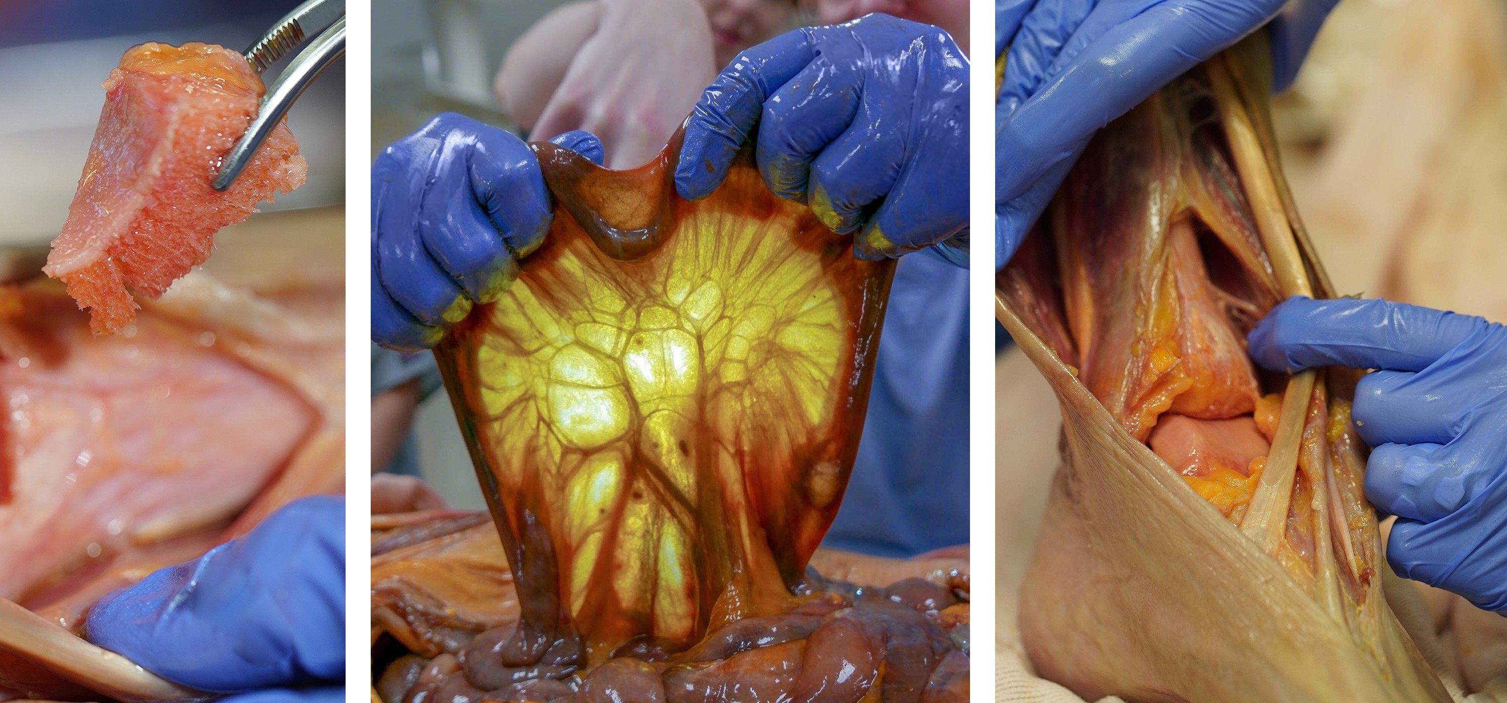
Tom Quisenaerts (Belgium) and Francis Van Glabbeek (Belgium)
Dissecting Two Arms: One live, and one prepared, following the 2006 Groningen University Study of Rembrandt’s The Anatomy Lesson of Dr Nicolaes Tulp
This project investigates the anatomical accuracy and perspectival construction of the dissected arm depicted in The Anatomy Lesson of Dr. Nicolaes Tulp by Rembrandt. Francis and Tom will dissect two left male arms under different conditions to explore both the preparatory and performative dimensions of anatomical demonstration.
One embalmed left male arm will be dissected in advance in the laboratory. During this procedure, the limb will be positioned according to the posture attributed to Dr. Tulp in the painting, with particular attention to reconstructing the vantage point from which the composition appears to have been created. Systematic photographic documentation will be undertaken from this reconstructed painter’s perspective to assess whether the anatomical structures represented correspond accurately to observable morphology.
A second left arm, preserved fresh-frozen, will be dissected live on site at the conference, echoing the historical setting of public anatomical lessons.
By combining laboratory-based reconstruction with live dissection, this study evaluates the interplay between anatomical fidelity, artistic composition, and viewpoint. The project contributes to ongoing discussions at the intersection of art history and anatomy, offering an empirically grounded reassessment of one of the most iconic representations of dissection in early modern Europe.
Biography
Francis Van Glabbeek is Professor at the Faculty of Medicine and Health Sciences of the University of Antwerp and an orthopedic surgeon at Antwerp University Hospital, where he serves as Vice-Chair of the Department of Orthopedics and Traumatology. Within the medical curriculum, he teaches musculoskeletal anatomy and contributes to courses on the history of medicine.
He has a long-standing interest in medical antiquarian books and has assembled a collection of more than one thousand works, including original publications by Andreas Vesalius, Govert Bidloo, Philipp Verheyen and Jan Palfijn. His medical-historical research focuses on lesser-known sixteenth-century healthcare practitioners, aiming to bring renewed attention to their lives and contributions.
He collaborates closely with historians and physicians including Maurits Biesbrouck, Theodoor Goddeeris and Omer Steeno. This work has fostered a broad international network of colleagues and friends among historians, physicians, bibliophiles and museum curators, including Robert Van Hee, Vivian Nutton, Monique Kornell, Daniel Margócsy, Stephen Joffe, Jacalyn Duffin and Jacqueline Vons.
His recent publications include Myology before Vesalius: Giovanni Battista Canani (1515–1579) (2022), Remacle and Gilbert Fusch (2024) and Jodocus Lommius and Anna van Egmond (2025). A forthcoming study on Balduinus Ronsseus is expected in 2027.
Tom Quisenaerts obtained his M.D. from the University of Antwerp and is currently pursuing his residency and PhD research in Plastic and Reconstructive Surgery. His research interests focus on breast cancer, reconstructive surgery, and medical statistics, as well as the medical humanities, particularly the intersections between medicine, art and history.
Through his involvement with BIOMAB, he has participated in several live anatomical dissections together with Francis Van Glabbeek. These demonstrations were organised for interdisciplinary audiences, bringing together artists and medical professionals to explore anatomical knowledge from both scientific and artistic perspectives.
Beatrijs Wolters van der Wey (Belgium)
Under the Knife. The Anatomy Lesson of Dr Joannes van Buyten (1648) as a Unique Source of Information about Surgery Lessons in Early Modern Antwerp
The Anatomy Lesson of Dr Joannes van Buyten, now preserved in the collection of the Royal Museum of Fine Arts Antwerp (KMSKA, inv. no. 610), is an impressive group portrait depicting surgeons attending an anatomy lesson. Commissioned in 1648 by the Antwerp surgeons’ guild, the painting was executed by the portrait specialist Frans Denys. It is the only known representation of this subject produced in the Southern Netherlands.
Beyond its art-historical significance, the work holds exceptional value for the history of medicine. Denys’s painting constitutes a major visual testimony to the practice of anatomy lessons within the framework of surgical apprenticeship in seventeenth-century Antwerp. Nineteen surgeons are portrayed within a real anatomy theatre, whose octagonal structure shapes the composition and spatial organization of the scene. The architectural setting is likely a faithful representation of Antwerp’s anatomy theatre.
As an iconographic source, when considered alongside seventeenth- and eighteenth-century archival documents, the painting offers important insight into the composition and functioning of the surgeons’ guild, as well as into the organization and conditions of surgical instruction in Early Modern Antwerp.
Biography
Beatrijs Wolters van der Wey studied Classical Philology, Art History and Healthcare Management and Policy at the University of Antwerp (UFSIA) and at KU Leuven, where she obtained her PhD in Art History in 2012. Her doctoral dissertation on civic group portraits in the historic Duchy of Brabant in the early modern period was awarded the Erik Duverger Prize by the Royal Flemish Academy of Belgium for Science and the Arts in 2013.
From 2004 to 2018 she was attached to the Documentation Department of the Royal Institute for Cultural Heritage (KIK-IRPA) in Brussels and held various teaching positions. She currently teaches at the Kunsthistorisch Instituut Antwerpen vzw and serves as Chair of its Executive Board.
As an independent scholar, she publishes on sixteenth- to eighteenth-century Flemish painting, with particular attention to the contextualisation of works of art and to material culture in general.
Figure. Frans Denys, De anatomische les van Dr. Joannes van Buyten, 1648-49 (Collectie KMSKA - Vlaamse Gemeenschap)

Click the image to view it in full.
DAY 2 - 8 May 2026
Grauwzusters Convention Center, Lange Sint-Annastraat 7, Antwerp
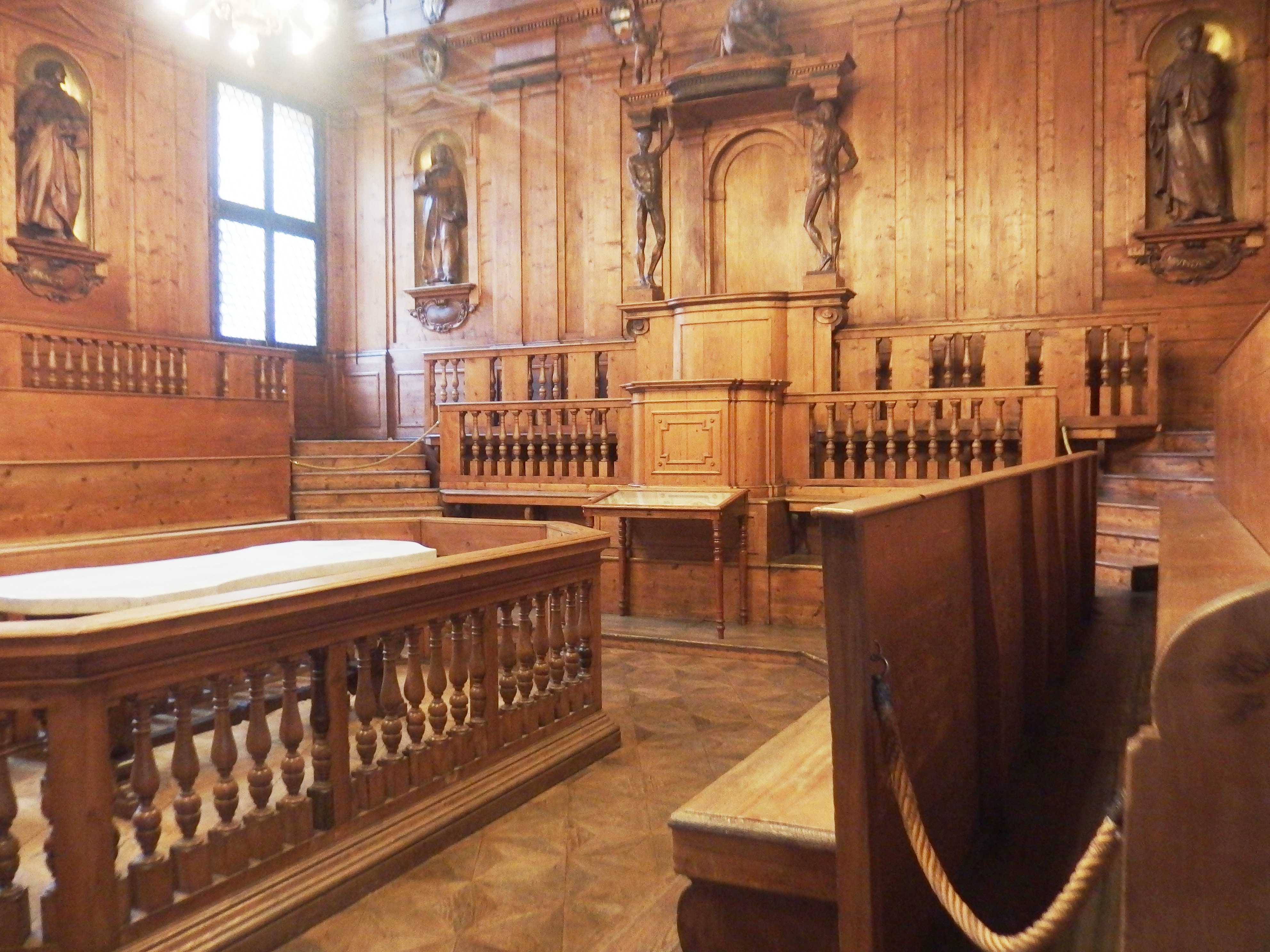
Christine Beese (Germany)
Scientific Instruments? On the Use of Sculptures in Anatomical Theatres
In anatomical theatres, bodies and bodily representations converge, differing in materiality and degrees of vitality. On the dissection table lies the freshly deceased body; around it assemble observers of various ages and levels of expertise. Along the walls and in vitrines, anatomical specimens, wax models, and sculptures in wood, plaster, and marble are displayed. Pictorial representations appear in handbooks, on charts, and within the decorative programs of the space, including fresco cycles.
From a modern perspective, these bodies often seem to embody opposing states—dead or alive, natural or artificial—and to function either as primary research objects or as secondary models. This paper examines the historical meanings and epistemic functions attributed to such bodily representations within processes of knowledge production. Particular attention is given to sculptures in wood, stone, and plaster, which are frequently interpreted today as artworks external to scientific practice. Drawing on the theoretical frameworks of Bruno Latour and Hans-Jorg Rheinberger, the paper argues that these objects formed integral components of an “experimental system.” Spatially realized within the “laboratory” of the anatomical theatre, they actively participated in the production of knowledge.
Focusing on the anatomical theatres of Bologna (seventeenth century), Ferrara (eighteenth century), and Boston (twentieth century), the paper demonstrates how shifting conceptions of art, nature, and science shaped the roles and meanings attributed to sculpture in anatomical theatres across different historical contexts.
Biography
Christine Beese is a tenure-track Professor of Architectural History at the Institute of Art History at Ruhr University Bochum. She leads an Emmy Noether Research Group funded by the German Research Foundation (DFG), dedicated to examining anatomical theatres as architectural spaces and their role in shaping a scientific public sphere in the early modern period. Her research situates these structures at the intersection of art, nature, and science, exploring how built environments contributed to knowledge production and visualization.
Beese’s scholarship focuses on the spatial, visual, and epistemic dimensions of early modern science. In “Between Allegory and Instrument” (2025), she analyzes the seventeenth-century London anatomy theatres designed by Inigo Jones and Robert Hooke as sites of visualization. Her article in Privacy Studies Journal (2025) investigates the anatomy towers of Göttingen and Jena, addressing questions of seclusion, spatial isolation, and knowledge-making in the eighteenth century. In kunsttexte.de (2023), she examines anatomical theatres as objects of art historical inquiry, reflecting on their dual function as imaginative spaces and “seeing machines.”
Louis Caron (USA)
Audiences and Anatomical Learning after Vesalius’s Fabrica
Audiences played a pivotal role in shaping anatomical learning in Early Modern Europe, particularly during Andreas Vesalius’s demonstrations at Padua and Bologna.
While recent scholarship has illuminated some aspects of this relationship, significant gaps remain, largely due to fragmentary evidence regarding what audiences actually experienced and how they engaged with public and private anatomical inquiry.
This paper surveys the existing evidence on audiences, exploring their diverse roles in structuring anatomical demonstrations and their influence on the dissemination of knowledge. Caron argues that anatomical learning cannot be understood solely through printed texts or the work of individual scholars; it was also shaped by the expectations, expertise, and participation of live audiences.
By examining how audiences familiar with Vesalius’s Fabrica influenced the conduct and presentation of dissections, this study highlights the dynamic interplay between printed knowledge and performative practice, showing that anatomical understanding was as much a social as an intellectual process.
Biography
Louis Caron has a deep passion for early modern European history and completed his graduate studies at Cambridge University. His research focuses on the history of European thought, with particular interest in the intersections of medicine, science (or natural philosophy), and political and religious ideas.
He currently teaches at the Crane Country Day School in the United States, helping students think critically about the past and engage with political concepts. Alongside teaching, he continues to research and publish on European intellectual history and considers himself privileged to be invited to speak at this conference.
Figure. Vesalius Teaching, from De humani corporis fabrica, 1543.
Theo Dirix (Belgium)
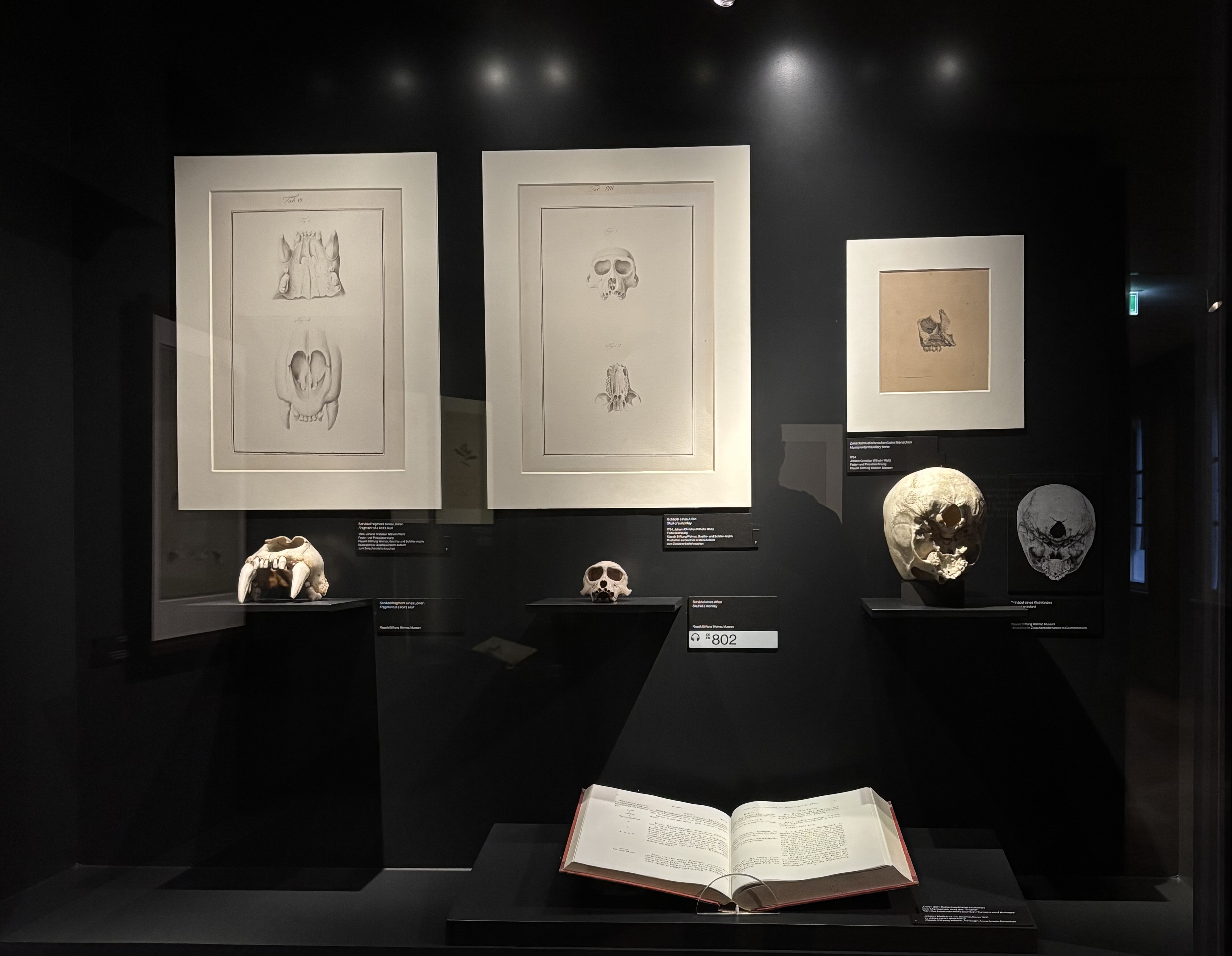
From obscura sutura to the Archetype: Vesalius, Goethe, and the theatrical staging of anatomical knowledge
This lecture examines how Johann Wolfgang von Goethe used the debate on the human intermaxillary bone to articulate his broader idea of the Urform (archetype). Goethe’s claim that humans possess a premaxillary bone was not only an anatomical intervention but also a gesture aimed at demonstrating morphological continuity between humans and animals. The episode highlights the complex interplay between visual observation, aesthetic judgment, and prior conviction in the making of anatomical authority.
The argument develops along three lines. First, Goethe’s anatomical claim is situated within contemporary debates by comparing his observations with those of leading anatomists of the time. Second, the Urform is discussed as a guiding concept that shaped what could be seen and recognized in the skeleton, giving coherence to otherwise ambiguous signs. Third, the case is placed within the broader context of anatomical practice as a theatrical space, in which demonstrating, drawing, and publishing worked together to stabilize uncertain knowledge. Vesalius’s notion of an obscura sutura serves as an early example of such ambiguity and illustrates how anatomical material itself invites multiple interpretations.
The lecture concludes that Goethe’s premaxillary argument shows how philosophical and aesthetic ideas influence what counts as anatomical observation. More broadly, it supports the conference’s central claim that anatomical knowledge is produced through acts of presentation and persuasion, in which style and representation are integral to scientific credibility.
Biography
Theo Dirix is a Flemish author, known mainly for his writings on literature and Andreas Vesalius, who now specialises in cross-disciplinary lecture-performances that merge text, music, and visual elements.
After a nomadic diplomatic life, he has returned to his first love: theatre. His recent works, A Necromantic Night, Vesalian Landscapes, and When AI Kills Faust, combine spoken word and live music to offer a sharp, poetic reflection on contemporary culture.
Figure. Photo by the author
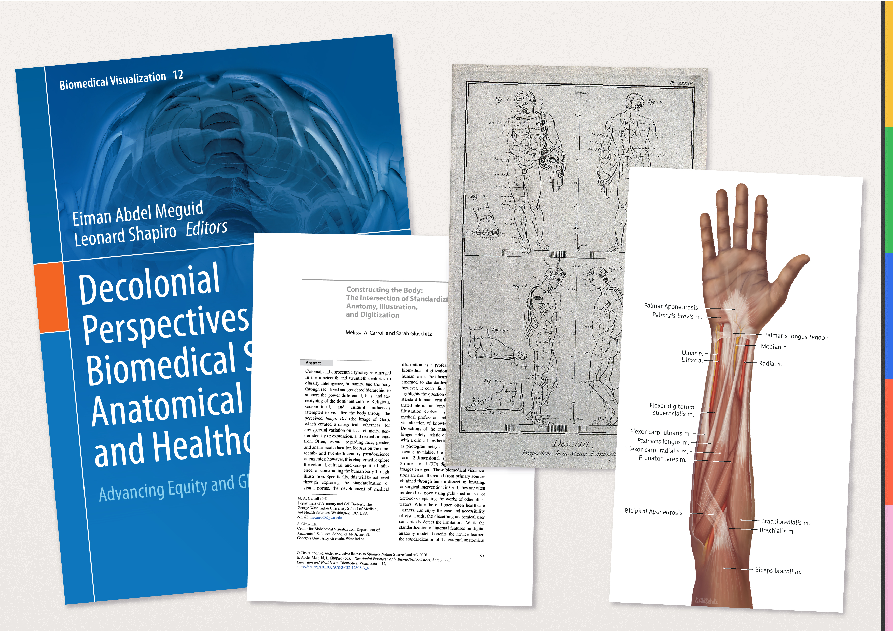
Sarah Gluschitz (Grenada)
Constructing the Body - The intersection of Standardizing anatomy, Illustration, and Digitization
Co-author Melissa A. Carroll
Abstract
Colonial and Eurocentric typologies emerged in the 19th and 20th centuries to classify intelligence, humanity, and the body through racialized and gendered hierarchies to support the power differential, bias, and stereotyping of the dominant culture. Religious, sociopolitical, and cultural influences attempted to visualize the body through the perceived Imago Dei (the image of God), which created a categorical ‘otherness’ for any spectral variation on race, ethnicity, gender identity or expression, and sexual orientation. Often, research regarding race, gender, and anatomical education focuses on the 19th and 20th-century pseudoscience of eugenics; however, this chapter will explore the colonial, cultural, and sociopolitical influences on constructing the human body through illustration. Specifically, this will be achieved through exploring the standardization of visual norms, the development of medical illustration as a profession, and the current biomedical digitization of the anatomical human form. The illustrated ‘homo perfectus’ emerged to standardize anatomical images; however, it contradicts human diversity and highlights the question of who determines the standard human form that contains the illustrated internal anatomy. Professional medical illustration evolved symbiotically with the medical profession and provided significant visualization of knowledge within the field. Depictions of the anatomical image are no longer solely artistic carbon dust renderings with a clinical aesthetic. As technology such as photogrammetry and stereoscopic images became available, the opportunity to transform 2-dimensional (2D) renderings into 3-dimensional (3D) digitized manipulatable images emerged. These biomedical visualizations are not all created from primary sources obtained through human dissection, imaging, or surgical intervention; instead, they are often rendered de novo using published atlases or textbooks depicting the works of other illustrators. While the end user, often healthcare learners, can enjoy the ease and accessibility of visual aids, the discerning anatomical user can quickly detect the limitations. While the standardization of internal features on digital anatomy models benefits the novice learner, the standardization of the external anatomical features removes humanity, erasing human diversity and variation from anatomy. Lacking access to primary sources (i.e., anatomical dissection) may limit the diversification of images constructed by skilled illustrators. The active and intentional encouragement of collaboration between trained anatomists and illustrators to discuss differing perspectives can significantly contribute to re-centering, reclaiming and decolonizing the standardized anatomical image.
Abbreviated description
This talk explores the religious, sociopolitical, and cultural influences which have attempted to visualize the human body, enforced through colonial and eurocentric typologies. Unravelling their influence on constructing the image of the human body through illustrations, specifically the standardization of visual norms, the development of medical illustration as a profession and the current biomedical digitization of the anatomical human form. Culminating in re-centering, reclaiming and decolonizing the standard anatomical image.
Keywords: Colonialism, Education, Enlightenment, Proportions, Visualization
Biography
Sarah Gluschitz is a Senior Medical and Scientific Illustrator and a Board-Certified Medical Illustrator (BCMI) and currently serves as a Full-Time Instructor in Medical Illustration in the Department of Anatomical Sciences at St. George’s University.
Since joining St. George’s University in 2018—first as a Demonstrator and later as an Instructor—Gluschitz has combined anatomical teaching with critical inquiry into representation in medical imagery. Gluschitz work challenges conventional visual models in medicine, particularly in relation to menstrual cycle illustration, encouraging more inclusive and conceptually nuanced approaches to visual communication in the health sciences.
Gluschitz holds an MA in Scientific Illustration from ZUYD University of Applied Sciences and Maastricht University, and a BA in Interactive/Media/Design from the Royal Academy of Art in the Netherlands and is an active member within the Association of Medical Illustrators, serving as Co-chair of the Diversity Committee and as a member of the Mentorship Committee.
Figure: Publication and artwork by the author, HR with AI (Ed).
Alison Klairmont Lingo (USA)
The Myth of the Wandering Womb and Its Metaphors in the Work of Ambroise Paré and Louise Bourgeois After Vesalius’s De humanis corporis fabrica (1543)
For much of the early modern period, the pregnant belly remained an opaque mystery—“truly transparent only in the vision of God.” Its invisibility stimulated a proliferation of texts promising to unveil the so-called “secrets of women,” a trope that spread widely across medical and popular literature and encouraged both anatomical inquiry and the dissection of female bodies. Notably, the first human internal organ to be represented in an Italian work on the basis of personal observation was the uterus, underscoring the intensity of male curiosity about female reproductive power. This curiosity contributed to the anatomical transformations culminating in De humani corporis fabrica (1543) by Andreas Vesalius. While some physicians advanced more positive interpretations of female anatomy, the terminology applied to women’s bodies ranged from neutral to pejorative and even pornographic. Meanwhile, the enduring authority of ancient thinkers—Plato, Aristotle, and Galen—as well as the Hippocratic corpus and popular beliefs about the uterus, made it difficult to disentangle empirical observation from inherited myth.
Among the most persistent of these myths was that of the wandering womb, believed to cause a wide range of physical and psychological disorders. Even the circulation of accurate anatomical illustrations demonstrating the uterus to be physically anchored did not entirely dispel the notion that it could move within the body. Public and private dissections of female cadavers had limited impact on entrenched assumptions.
Medical metaphors describing women’s “nature” further reinforced the myth. This paper examines how such metaphors operated in the writings of Louise Bourgeois, Ambroise Pare, and Jean Liebault. It argues that Bourgeois—the first woman to publish a medical text in France—challenged prevailing figurative language surrounding the womb, thereby offering a more nuanced understanding of female anatomy and destabilizing a long-standing medical myth.
Biography
Alison Klairmont Lingo is a Research Associate in the Department of History at the University of California Berkeley, where she has been affiliated since 2014. She earned her Ph.D. in early modern history at Berkeley, specializing in gender, medicine, and the cultural history of early modern France. Her work explores the intersections of medical practice, women’s authorship, and material culture in the seventeenth century.
Lingo is best known for her critical edition and translation of Louise Bourgeois, Midwife to the Queen of France: Diverse Observations (1626 edition) (Toronto, 2017). This landmark publication—the first complete English translation of Observations diverses and the first critical edition in any language—received the Josephine Roberts Award for the best scholarly edition in early modern women’s and gender studies. The volume restores the voice of Louise Bourgeois, midwife to the French queen, and highlights her central role in the history of obstetrics and women’s medical writing.
Her recent publications include “Natalie Zemon Davis and the Origins of Women’s History at Berkeley: A Former Student’s Perspective,” published in Archiv für Reformationsgeschichte / Archive for Reformation History (2025), and “The Material Culture of the Birthing Room in Seventeenth-Century France: The Hand and Other Instruments,” featured in Maternal Materialities (Brepols, 2024). Through her scholarship, Lingo continues to advance the study of gender and medical knowledge in early modern Europe.
Monique Kornell (USA)
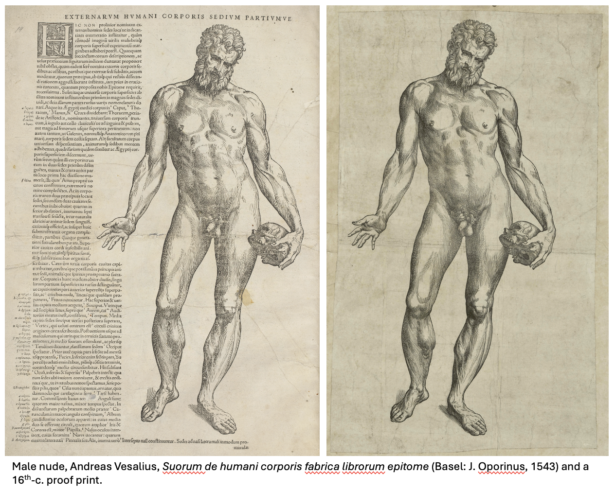
Vesalius’s Epitome (1543): The Illustrations and an Early Proof Impression
Andreas Vesalius’s Epitome, a brief illustrated summary of anatomy, is described by the author as a compendium of the books of the Fabrica in two parts. These are comprised of a short text and a set of eleven woodcut illustrations, two leaves of which were meant to be cut-up with the resulting pieces glued together to create flap-anatomy manikins of both genders. The Epitome’s reader is invited to start either with the text or the illustrations. This choice is an indication of the autonomous nature of the text and images, for unlike the Fabrica, there are no references to the illustrations in the Epitome text. The text was repeatedly copied in the 16th and 17th centuries, often packaged with a selection of illustrations after the Fabrica and the male and female nudes of the Epitome. This talk will consider the illustrations of the Epitome and the history of the woodblocks in light of a newly identified proof impression of the Epitome’s male nude figure, printed without its surrounding text. A consideration of its provenance and an analysis of the wear of the related woodblock through later printings establishes that this proof impression is an early one, possibly one made for Vesalius himself in the course of the preparation of the Epitome. Vesalius worked from proof prints as well as drawings when writing about the illustrations of the Fabrica and the Epitome, and a set of proofs interleaved with the woodblocks was sent as a guide to his printer, Johannes Oporinus in Basel, with a request that he follow them closely.
Biography
Monique Kornell is an art historian whose scholarship explores the dynamic relationship between art, anatomy, and the history of medicine from the Renaissance to the Enlightenment. She is Honorary Director of the Program in the History of Medicine at the Center for the Arts and Humanities in Medicine at Cedars-Sinai Medical Center, where she also serves as Visiting Associate Professor in the Department of Biomedical Sciences and convenes the 2025–26 seminar series The Book in the History of Medicine. She earned her Ph.D. from the Warburg Institute, University of London, following graduate study at the Courtauld Institute of Art and a B.A. with distinction from the University of Toronto; she also completed anatomy training for artists at University College London.
Kornell has held fellowships and research appointments at the Wellcome Institute for the History of Medicine and the University of California, Los Angeles, and served as Guest Curator at the Getty Research Institute. There she curated Flesh and Bones: The Art of Anatomy (2022), with a new iteration forthcoming at the ArtScience Museum, Singapore (2026). A leading authority on Andreas Vesalius, her publications examine print culture, illustration, and anatomical knowledge, illuminating how books and images shaped medical understanding across early modern Europe.
Figure. Male nude, Andreas Vesalius, Suorum de humani corporis fabrica librorum epitome (Basel: J. Oporinus, 1543) and a 16th-c. proof print.
Daniel Margócsy (UK)
Vesalian knowledge spread throughout Asia into Japan
This lecture explores the circulation of Vesalian imagery in Asia during the early modern period, with particular attention to the networks of the Dutch East India Company (VOC). By the late seventeenth century, books and illustrations derived from De humani corporis fabrica had reached various parts of Asia, where they intersected with local cultural, religious, and medical traditions.
Focusing on the motivations of VOC officials, the talk examines why and how they engaged with Vesalian anatomical knowledge, including its practical, intellectual, and symbolic uses. It also considers how these images may have been received by local audiences, proposing hypotheses about their interpretation and adaptation in non-European settings.
Drawing on case studies from the Indonesian archipelago, Japan, and South India, the lecture highlights the complex exchanges that shaped the transmission and transformation of anatomical knowledge across cultures.
Biography
Dániel Margócsy is Professor of the History of Science, Technology and Medicine at University of Cambridge. His research explores the global circulation of knowledge in the early modern period, with particular emphasis on visual culture, natural history, and the relationship between science, commerce, and colonialism.
Margócsy studied at Harvard University, where he completed his PhD in the History of Science, following earlier studies at Stanford University and University College Utrecht. Before joining Cambridge, he held academic positions at the City University of New York and was a postdoctoral fellow at Northwestern University.
His publications include the award-winning Commercial Visions: Science, Trade and Visual Culture in the Dutch Golden Age and The Fabrica of Andreas Vesalius: A Worldwide Descriptive Census. His work has received numerous distinctions, including a fellowship from the John Simon Guggenheim Memorial Foundation and the Neu-Whitrow Prize.
Margócsy’s current research investigates the movement of knowledge through global trading networks, especially within the Dutch East India Company world. In 2024, he was appointed Council Member of the British Society for the History of Science and became an Ordinary Fellow of the Center for the Study of Medicine and the Body in the Renaissance.
Vivian Nutton (UK)
Subject to unforeseen circumstances
Kevin Petti (Italy)
Connecting Art and Anatomy in Italy
The Italian peninsula offers a unique story, as its medieval universities established the study of human anatomy for physicians, later serving as the cradle of the Renaissance. The profound connection between art and science in Italy is beautifully illustrated by Michelangelo: the wooden crucifix he carved in gratitude for secret access to corpses from a convent hospital still hangs in the Basilica of Santo Spirito in Florence.
This talk explores the nexus between art and science in Italy, examining the history of anatomy education in the first universities, discussing how Renaissance masters clandestinely conducted dissections to enhance their art, and considering how that knowledge influenced some of the world’s greatest artists, including Leonardo da Vinci, Michelangelo, and Raphael.
Participants will reframe Renaissance masterpieces within the context of human dissection, analyze the influence of Renaissance art on early anatomy texts, and compare and contrast the accuracy of early anatomy texts with the anatomical representations of select Renaissance masters.
Biography
Kevin Petti, PhD is a dual United States of America/Italian citizen, college professor, textbook coauthor, and president-emeritus of the Human Anatomy and Physiology Society. He teaches anatomy and physiology, human dissection, and health and human behavior at San Diego Miramar College, and leads study-abroad programs across Italy and Europe through the Anatomia Italiana program he founded in 2012, exploring the origins of anatomy as a science and its influence on Renaissance masters. His students include anatomy professors, physicians pursuing continuing education, and undergraduates from San Diego State University.
Petti is an invited speaker on the connection between art and anatomy in Greco-Roman, Medieval, and Renaissance Italy at international conferences, museums, Italian-American groups, and universities throughout North America and Europe. He has been invited by the Italian government to speak at their Cultural Institutes in the United States of America and at the Italian Embassy in Washington, D.C. The University of Palermo, Sicily, hosted him as a guest lecturer for its 210th-anniversary seminar series, and he was featured in the eight-part Chinese Central Television (CCTV) documentary, 200 Years of Surgery.
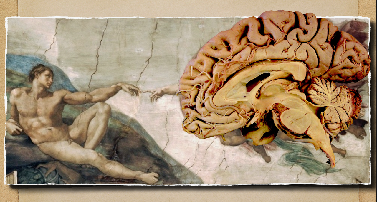
Figure. The Creation of Adam, Michelangelo, c. 1512, fresco, Sistine Chapel. Author’s collage incorporating brain imagery.
Gitte Samoy (Belgium)
Itinerant Bodies: Anatomy and Medicine at the Fairground, Nineteenth–Twentieth Centuries
One of the staple attractions of the nineteenth-century funfair was the popular anatomy museum. These venues revealed the secrets of human anatomy alongside the grotesque sights of deviant and sick bodies. This paper presents a short history of these museums and the objects and people behind them. Focusing on the performative and material dynamics of these exhibitions, I argue that they played an important role in the broader circulation of medical knowledge.
While spectacle and artistry were key to attracting visitors, these itinerant cabinets were often assembled from the same makers and objects as institutional collections. Wax modellers, in particular, appear as central figures in producing and mediating anatomical objects, bridging professional medicine and public entertainment. The presence of anatomical museums at the fairground also signalled the event’s potential as a civilized and morally acceptable form of leisure.
As academic medicine professionalized, tensions between the spectacle of the funfair and the moral contemplation encouraged by these cabinets grew, ultimately contributing to their decline. Although this history formally ended by the First World War, its legacies persisted well into the twentieth century.
Biography
Gitte Samoy is a PhD researcher at the University of Antwerp, where she is a member of the Arts & Media Archaeology team at the Antwerp Research Institute for the Arts and participates in the ERC-funded project Science at the Fair (www.scifair.eu). She holds a Master’s in History from the Catholic University of Leuven and a Master’s in African Studies from the University of Ghent.
Her doctoral research, titled Spectacular Bodies: Performing Anatomy, Medicine and Anthropology, examines the circulation of knowledge about the body at funfairs in Northwestern Europe during the nineteenth and early twentieth centuries. She focuses on the role of materiality and performative strategies in shaping ideas about health and disease, disability, gender, class, and race, and explores how these notions were reinforced or challenged through the fairground experience.
Figure. Leaflet 'Grand museum anatomique Dr. Spitzner du Chateau-d'Eau de Paris', ca. 1887, University library Ghent.
Guido Sold (Germany)
In the footsteps of his forefathers in Wesel: Andreas Vesalius’s origins
This talk provides an overview of the ancestral background of the Vesalian family, highlighting major figures and correcting misconceptions associated with the name, known not only in the sixteenth but also in the fifteenth century.
First, the family’s original Rhenish name—Wytinc(k), also Wijtink, Vitinch, Viting, and variants—likely does not refer to the whiting recorded in the kitchen books of Katherina van Kleef (1417–1476), a contemporary of Jan Wytinck (1400/1401–1476). The name can be traced back to the 13th century with Harmannus filius Witen (1289) and successive members serving as schepen and burmeister throughout the 14th century. The suffix -in/-ing/-inc(k) signifies “son of,” and heraldic evidence links the family to the city of Wesel and, after 1400, to the house of Cleves.
Second, despite Vesalius stating in his 1546 China Root Letter that his great-grandfather was Petrus, archival sources confirm that Everard was the true ancestor, father of Jan Wytinck, doctor in medicinis et expertus in astronomia, and Hermann, merchant in Wesel. No Peter appears in the lineage from 1289 to 1564.
Third, the Rhine-Meuse region remained linguistically undivided into the early modern period, so Jan Wytinck could communicate easily in Brabant. Combined with Wesel’s prominent role in the Hanseatic League, this positioned him in a socially and politically advantageous environment.
Biography
Guido Sold studied medicine in Heidelberg—where he was influenced by Heinrich Schipperges—and in Sheffield, after completing his schooling in Speyer with a focus on Latin, ancient Greek, and French. He earned a doctorate in biochemistry and began his clinical career in Kempten (Germany) and Brunssum (Netherlands).
In 1976, he joined the University of Göttingen, specializing in noninvasive cardiology. His work led to the publication of a textbook on two-dimensional echocardiography, including Doppler techniques. In 1992, he became head cardiologist and angiologist at the Evangelisches Krankenhaus in Wesel, a position he held until his retirement in 2014, the 500th anniversary of the birth of Andreas Vesalius.
Alongside medicine, Sold has been active in early music, founding the ensemble Convivium musicale, Wesel and serving as president of the Deutsche Clavichord Societät (2016–2022). Since 2020/21, he has been a guest student at Ruhr University Bochum, focusing on late medieval and early modern history, particularly the Vesalian family.
Together with friends and like-minded people he founded a “Forum Vesalius/Wytinck” in Wesel.
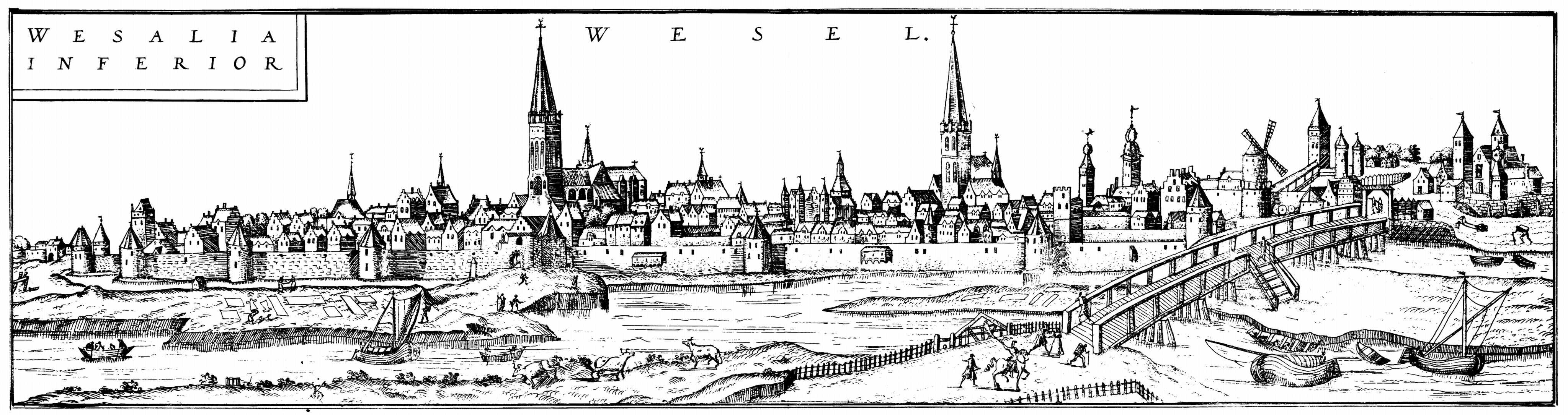
Figure. Engraving "Wesalia Inferior" (referring to Wesel on the Lower Rhine), created by Franz Hogenberg and published in the seminal town atlas Civitates Orbis Terrarum (Cities of the World) around 1572.
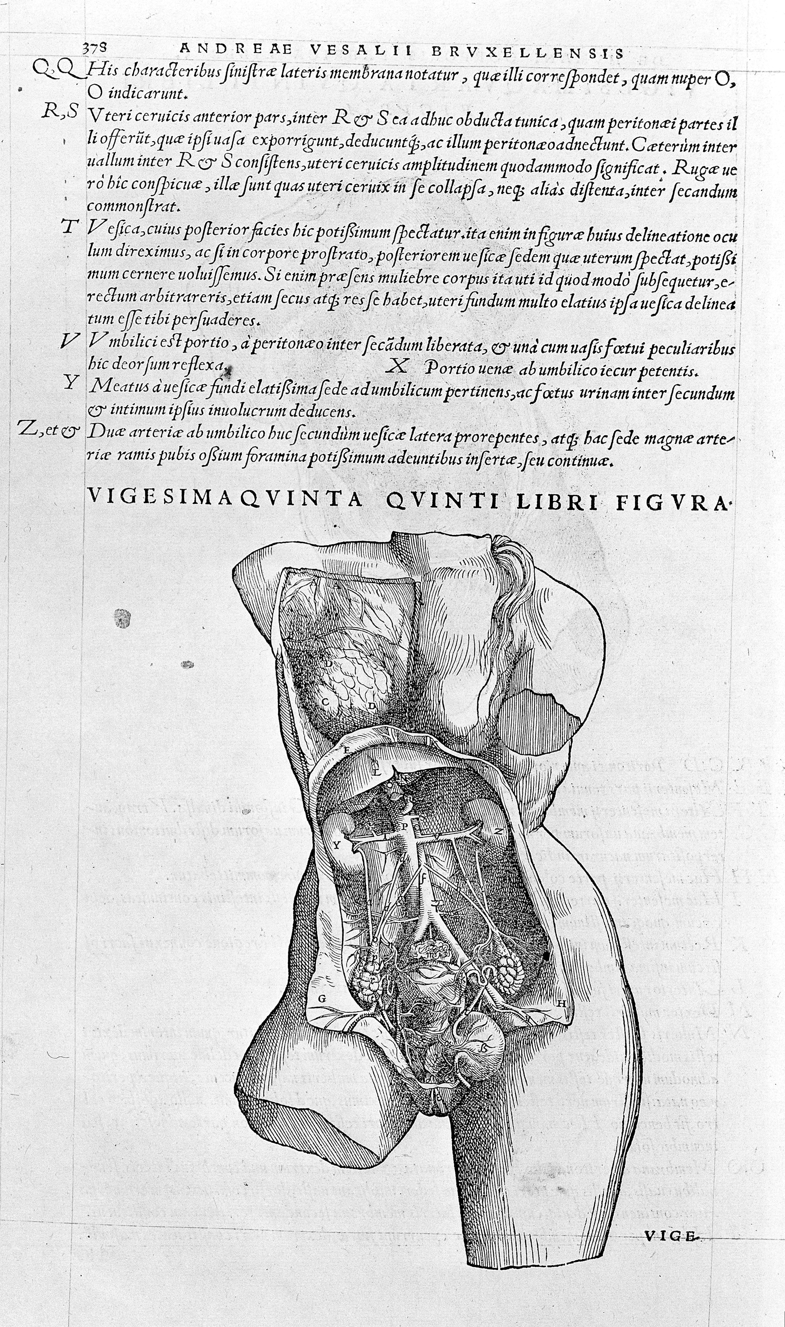
Jacqueline Vons (France)
A New Reading of Vesalius’s Fabrica
How can we move beyond the traditional descriptive approach to anatomy? By focusing on the digestive organs in Book V and the respiratory organs in Book VI of the Fabrica, Vesalius not only highlights the interrelationships between organs, but also offers what can be considered the first modern “manual” of dissection. This approach allowed his students and successors to learn proper dissection techniques, replicate them accurately, and thereby ensure the systematic teaching of anatomy in universities.
(Presentation in French – PowerPoint in English)
Biography
Jacqueline Vons is Professor Emerita and Honorary HDR Researcher at the University of Tours, specializing in Latin and the history of medicine. She is the author of numerous books and articles on medical texts of the sixteenth and seventeenth centuries. Recent publications include Littérature et médecine. Les mots et les maux (with C. La Charité, RHLF, 2020); Dissection et représentation des muscles chez Vésale, Canano, Sagemolen (with F. Van Glabbeek), in Quatre atlas de myologie de Van Horne et Sagemolen (J.F. Vincent & I. Bonnard, eds., Univ. Paris Cité, 2022); and Vesalius and the Timaeus: The Anatomist’s Answer to the Philosopher, in The Legacy of Plato’s Timaeus (J. Prins & E. Thomas, eds., Brill, Leiden, 2025, pp. 365–385).
Together with Stéphane Velut, she published the first French edition and translation of Vesalius’s De humani corporis fabrica (available online). She serves as President of the Académie des sciences, arts et belles-lettres de Touraine and is Editor-in-Chief of Les carnets d’histoire de la médecine.
Figure. Vesalius Teaching, from De humani corporis fabrica, 1543.
Daniella Zaidman-Mauer (The Netherlands)
Vernacular Anatomy: A Sixteenth-Century Yiddish Translation of Vesalius’s Epitome
This presentation examines a previously understudied manuscript preserved in the University of Pennsylvania Libraries, Lawrence J. Schoenberg Collection (LJS 485): a Yiddish translation of Andreas Vesalius’s De humani corporis fabrica librorum epitome (1543). Written in Ashkenazi script, the manuscript provides rare evidence of Jewish engagement with Renaissance anatomical knowledge and of the transmission of Vesalian ideas beyond a Latin-literate readership.
The existence of such a vernacular translation raises important questions. Who produced this text, and for whom was it intended? What role could anatomical knowledge assume within Jewish communities, where dissection was prohibited by religious law? By situating this Yiddish Epitome within the broader landscape of vernacular medical translation, the paper contributes to current scholarship on the cross-cultural transmission of anatomical knowledge in early modern Europe.
Biography
Daniella Zaidman-Mauer is a scholar of Early Modern and Modern Yiddish literature and vernacular medical texts, focusing on how Jewish communities in Europe engaged with healing knowledge and epidemics.
She earned her PhD at the University of Amsterdam (Amsterdam School of Historical Studies) with a dissertation titled: Plague and Piety. Yiddish Medical Literature in Early Modern Europe.
She teaches at the University of Amsterdam, The Netherlands.
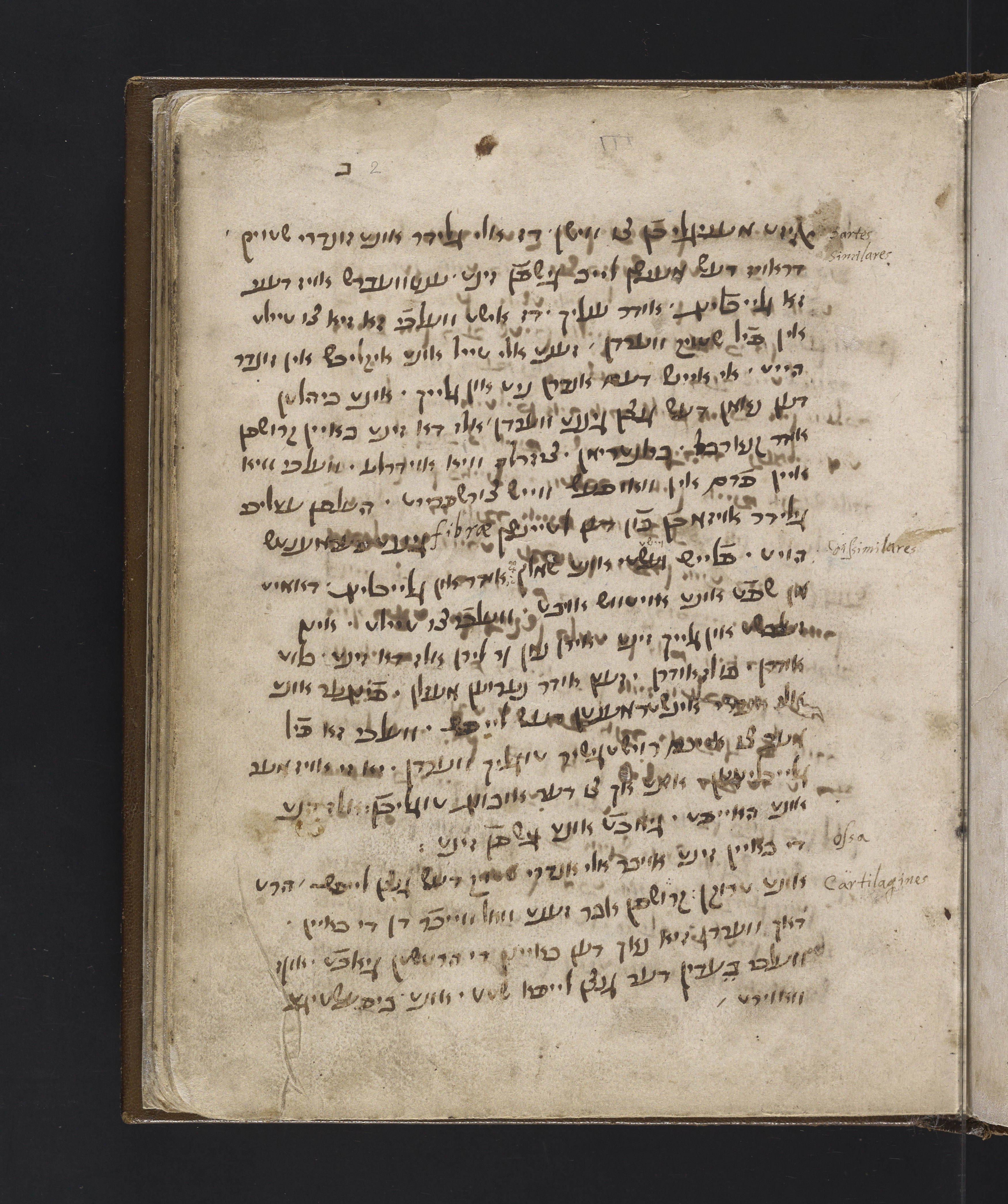
Figure. Scan of Yiddish ms., translation of Vesalius's Epitome (c.1590-5), MS 485, Lawrence J. Schoenberg Collection · University of Pennsylvania Libraries.

Click the image to view it in full.
DAY 3 - 9 May 2026
Lambotte Museum, Heilige Geeststraat 21, Antwerp