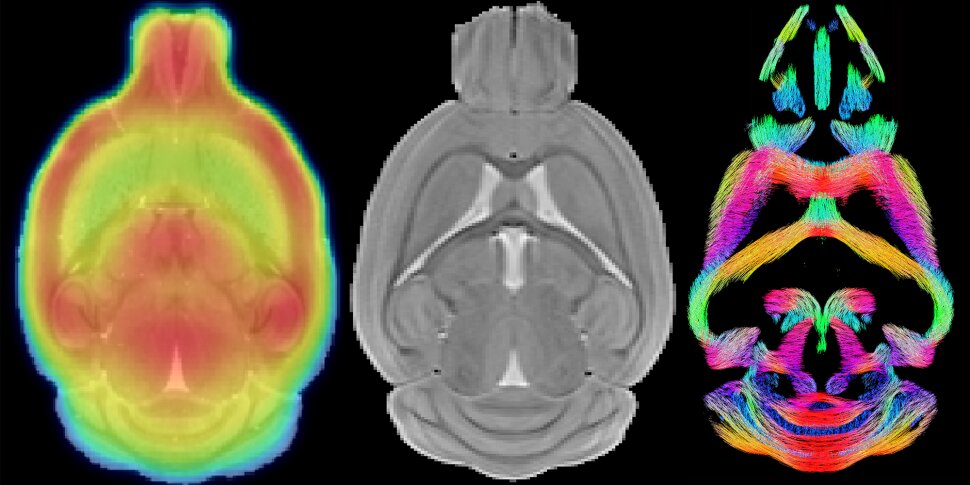
The Molecular Imaging Center Antwerp and Bio-Imaging Lab bring their small animal imaging expertise together in the MICA_BIL core facility
The core facility is actively supported by the University of Antwerp to facilitate collaborative projects between different academic research groups. The groups bring together key preclinical imaging expertise and facilities within the UAntwerp. The equipment makes it possible to make virtual sections through a living laboratory animal enabling to quantitatively monitor various anatomical, morphological, physiological and molecular processes over time in the same animal. In addition to the in‐vivo multi‐modal imaging systems, there is also access to a Bioluminescence/Fluorescence camera, animal monitoring, microsurgery and a radioprotected laboratory animal animalarium.
Objectives:
We are open to joint research collaborations within the University of Antwerp, national or international research institutes/universities and seek to provide services in PET/SPECT/MRI/MRS/BLI. These projects are typically consolidated within (double) PhD grants and any other type of mutual grants.
Collaborations are initiated by sending an informal pre-inquiry, briefly describing the aim of the study, requested experiments, species, number of animals (if applicable) to bio-imaginglab@uantwerpen.be.
Upon positive evaluation of the pre-inquiry, a detailed application by completing the ONLINE FORM is requested.
Available Infrastructure:
- 2 INVEON Siemens µPET/CT
- VECTor SPECT/CT
- On-site cyclotron and radiosynthesis
- 2 Bruker 7T MRI
- Bruker 9.4T MRI with cryo-coil
Readily Available MRI techniques:
- Structure analysis (volumetric and morphometry)
- Microstructural MRI (diffusion tensor, kurtosis, and fixel-based)
- Brain functional connectivity (Functional connectivity, CAPs and QPPs)
- Brain functional activity (stimulus-based and pharmaco-based fMRI)
- Susceptibility weighted Imaging (SWI and QSM)
- Cerebral Blood perfusion (CBF) and volume (CBV) using ASL
- Brain clearance (invasive and non-invasive glymphatic clearance)
- Manganese enhanced MRI (MEMRI)
- Magnetic resonance spectroscopy (MRS)
- Metabolic rate of O2 consumption; 31P MRS for energy metabolism
- Fluor-MRI; Magnetization transfer / Glutamate CEST
Readily available PET tracers
- Dopamine D1 Receptors (D1R) using [11C]SCH23390
- Dopamine D2/3 Receptors (D2/3R) using [11C]Raclopride
- Phosphodiesterase 10A (PDE10a) using [18F]MNI-659
- Pre-synaptic density (SV2A) using [18F]SynVesT-1
- Neuroinflammation (Translocator protein, TSPO) using [18F]PBR111
- Glucose metabolism using [18F]FDG
- mGluR5 using [11C]ABP688 or [18F]FPEB
- mHTT aggregates using [11C]CHDI-180R, [18F]CHDI-650, others…
- Beta-amyloid aggregates using [18F]AV-45 or [11C]PiB
