This page presents a number of highlights from the research conducted within the frame of this chair. The topics concern both instrumental development of imaging setups and applied research on specific works of art. The latter is mainly performed as a service to museums. This type of analysis is usually carried out to support an ongoing conservation treatment with insights into the artist's materials, techniques and degradation mechanisms. Nevertheless, for several Masters such Van Eyck, Van Gogh or Rubens the aim is to obtain a significant corpus of data that allows characterising the studio practice of these artists.
Girl with a pearl earring
Chemical Imaging on Works by Jan Van Eyck
The reverse side of the Ghent Altarpiece was fully examined during 2015 in the Museum of Fine Arts in Ghent. The experiments were performed in support of the first stage of the conservation treatment by the KIK-IRPA and had a direct impact on the treatment: the elemental images did not only reveal Van Eyck's hidden painting, they also showed that the original composition was in a relatively good preservation state. In this way, the MA-XRF scans underpinned the much debated decision to proceed with a complete removal of the overpainting.

In a second phase of the research, a number of exploratory MA-XRF scans were carried out on the frontside of the central panels, in the Cathedral of Saint Bavo in Ghent. Measurements were made on the panel of God the father and the central Lamb with the aim of assessing the extent of overpainting on the front side.In this way, the work duration and cost of the second phase could be estimated more accurately. In addition to that, the Portrait of Margaret, Virgin and Child with Canon van der Paele (Bruges), Saint Barbara and the Madonna at the Fountain (KMSKA) were scanned in support of the VERONA project of the KIK-IRPA
More on the Ghent Altarpiece
MA-XRF scans on works by Jheronimus Bosch
Two works by Bosch were analysed: Jerome in Prayer (MSK, Ghent) and Saint Christopher (Booijmans Van Beuningen, Rotterdam). At the time of the scans, both panel paintings were undergoing an extensive conservation treatment in preparation for the upcoming Bosch year and the associated exhibition in 's Hertogenbosch. Jerome in Prayer was treated in the studio of the KIK-IRPA by Aline Van Genbrugge while the Saint Chirstopher was treated in the Museum Gallery in Rotterdam by Annetje Boersam and Eva Van Zuien. For both paintings, the ensuing MA-XRF images reflected the complex genesis, the worn condition and an eventful conservation history.
The opportunity was seized to analyze other works attributed to Bosch in the Rotterdam museum as well, in particular The Wayfarer and two side wings from which the central panel is lost.

Collaboration with the Royal Museum of Fine Arts Antwerp (KMSKA)
J.D. De Heem – Still Life with Flowers and Insects
The central work of the 'Flower Power' exhibition (Rockoxhuis Nov 2015- March 2016) was scanned in support of the preceding conservation treatment. The flower still life was investigated in situ, both with XRF and XRD imaging. The measurements revealed the particular painting technique that was used by J.D. De Heem to realize the sparkling colours of his famous still lifes. Amongst other findings, the scans revealed that De Heem obtained an unprecedented depth of colors by applying a monochrome ellipse as underpainting for each flower. In addition to that, for the firs time the degradation products of orpiment were identified by MA-XRD imgaging in a completely non-destructive way. Orpiment is a yellow and highly toxic arsenic sulphide that was used by De Heem to paint the central yellow roses. Measurements showed that the pigment reacted with the existing white lead. As a result these central flowers lost their (optical) depth.

P.P. Rubens – Rockox Triptych
The Rockox triptych was treated in 2015-'16 by conservatorEva Van Zuien. MA-XRF scans were performed in order to obtain a better insight in the layer structure and use of materials, and to support the conservation treatment. Traditional X-Ray Radiographs of the wings proved difficult to read because the coats of arms of the donors were painted on the verso sides
Judith – Jan Masssijs
This 'topstuk' of the Antwerp school was treated in the course of 2015-'16. Previous research suggested that the appearence of the work had changed by discolored pigments and old restorations. MA-XRF scans allowed gaining a better understanding of the original appearance of the composition. The greyish robe of Judith appeared to contain smalt and therefore must have been (bright) blue in its original state. The cloth behind Judit is currently a dark, flat plane, but the resulting MA-XRF images demonstrated that the draping was green in color and must have exhibited much more depth and details. The clouds are brown, a degradation that requires further research on sample material, an interesting defect that was also found in other works of Jan Matsys.
Hans Memling - Christ with singing and music making angels
In the course of their long conservation treatment, these panels have been subject to a wide variety of chemical analysis. Although many questions concerning the painting technique and degradation phenomena were answered, these new insights established new questions . For that reason, additional MA-XRF and MA-rFTIR scans were performed.

Works by James Ensor
A series of works by Ensor was examined in the context of the ongoing Ensor research project led by Dr. Herwig Todts. The aim was to complement the existing knowledge about Ensor's use of pigments and to gain a better understanding of Ensor's reuse of canvases.
MA-XRF on Ceramics
Majolica: The Fall of Paul (MAS)
The Fall of Paul is one of the rare surviving 16th century majolica tableaus from the workshop of Guido Andries, the most important Antwerp majolica baker. This masterpiece was treated in the course of 2016 in preparation of a number of international exhibitions. The purpose of the imaging experiments was to assess the extent to which MA-XRF scanning can be employed to distinguish original tiles from 19th and 20th century inserts . The research is part of a larger inquiry to apply MA-XRF scanning on materials other than oil paintings.

MA-XRF Scanningof Stained Glass Windows
15th C stained glass window from Bruges depicting Saint George and Saint Michael
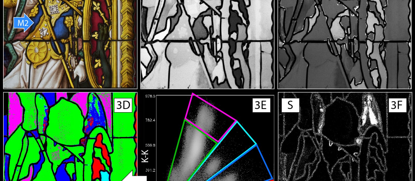
The applicability of MA-XRF scanning for investigating stained glass windows was assessed by measurements on a high-profile, well-studied late mediaeval panel. Although MA-XRF scanning did not permit an accurate quantification of components ,plotting the detected intensities of K versus Ca in a graph allowed to distinguish glass fragments of different compositional types within the same panel. In particular, clusters in the K/Ca correlation plot revealed the presence of two subtypes of potash glass and three subtypes of high lime low alkali glass. MA-XRF results proved consistent with previous quantitative SEM-EDX analysis on two samples and analytical-based theories on glass production in the Low Countries formulated in literature. Apart from identification of the chromophores responsible for the green, blue and red glass colors, contrasting the associated elemental distribution maps obtained on the exterior and interior side of the glass allowed discriminating between colored pot metal glass and multi-layered flashed glass.

The oldest leaded glass windows in Belgium
This series of round leaded glass windows (or occuli ) date from the 13th century and are the oldest preserved windows of Belgium. Early after their placement in the Cathedral of Saint Gudula in Brussels, the windows were covered by a stone wall, after which they remained hidden until they came to light during the latest restoration campaign of the cathedral. Although the glass is colorless, the glass pieces display various very light hues from light green to pale pink as a result of impurities in the raw materials,. Because of this variation, it is believed that the windows contain recycled glass, but untill now both visual study and analysis by means of SEM-EDX did not allow an accurate differentiation of the glass pieces. However, MA-XRF scanning permitted dividing the pieces into two groups, according to chemical trace elements detected in the glass.

Instrumental Development
Macro X-Ray Diffraction from the lab to the museum
The most innovative aspect of the existing macro X-ray fluorescence scanner (MA-XRF) is the fact that chemical data is translated into images that can be easyly interpreted by art historians and restorers. Apart from MA-XRF However, there are other analytical techniques that can provide additional information about the studio practice of artists. For that reason, the AXES group is working on the conversion of analytical techniques into mobile scanning devices. One of these techniques is X-ray diffraction, a method of analysis that is similar to XRF because both are based on the interaction of X-rays with matter. However, XRF characterizes the chemical elements in a material while XRD provides detailed information on crystal structures. A mobile MA-XRD scanner is promising for the technical examination of works of art for two reasons. First, the ensuing information is complementary with MA-XRF: XRD can identifies a number of paint components that that cannot be detected with MA-XRF. Secondly XRD is able to characterize degradation products. The latter offers an insight into the causes that lie at the basis of discolorations and defects and is therefore particularly valuable for the preservation of works of art. Up to now, this kind of research was only possible by analyzing samples extracted from the paint film, often in particle accelerators. The development of a mobile MA-XRD scanner is, in other words, an important step closer to completely non-destructive examination of paintings and/or will limit the extraction of paint samples.

Picture of the MA-XRF scanner (left) and the MA-XRD scanner (right) working side by side at the KMSKA
In the course of 2015, the development of the AXES 'macro-XRD scanner has progressed significantly. In first instance, a series of scans was carried out on so-called "mockups' - test paintings that imitate the structure of a real work. The next step consisted in the analysis of a number of historical paintings in our X-ray laboratory. Because both experiments proved successful, it was decided at the end of the year to move the XRD scanner to the museum depot of the Royal Museum of Fine Arts Antwerp to measure historical works in situ.
Scans on works by J.D. De Heem and Ensor appeared to meet the expectations. The results were communicated respectively during the Flower Power exhibition and a seminar on Ensor. Finally, a second campaign was conducted in the Kröller-Müller Museum on a late work by Van Gogh: Hay Stacks.
MA-XRF scans on Bosch: Saint Jerome in Prayer

Scans op een bloemstilleven van J.D. De Heem

Chemical imaging of stained-glass windows by means of macro X-ray fluorescence (MA-XRF) scanning