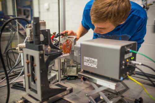
In-house MAXRPD scanner
Macroscopic X-ray powder diffraction scanning (MAXRPD) is a technique that allows to visualize the distribution of crystalline compounds in different types of flat macroscopic samples such as paintings or illuminated parchments. This is achieved by scanning the surface of the sample with a focused or collimated X-ray beam of (sub)mm dimensions and analyzing the diffracted radiation. This technique is non-invasive, non-destructive and can be performed in situ and can be performed in reflection and transmission mode. Simultaneously XRF collection is possible and a laser distance sensor ensures the scanner remains in focus during the scanning procedure.
The advantage of scanning in reflection mode is the high surface sensitivity due to the shallow angle configuration, typically 10°. In this manner MA-XRPD is not only a suitable technique to directly identify original pigments and distinguish them from non-original pigments, but it can also provide valuable insights into degradation processes occurring within the upper paint layers. The disadvantage is the slightly larger beam size of 0.8 x 0.2 mm2. By examining the sample in transmission mode, a smaller beam size can be achieved (0.2 x 0.2 mm2), but the obtained information will be representative for the bulk composition of the paint stratigraphy (e.g. thick paint and ground layers) instead. Two significant limitations for transmission MA-XRPD are that the object has to be moved relative to the equipment (e.g. large objects or paintings fixed to the wall cannot be scanned) and that the sample needs to be sufficiently thin for the X-rays to pass through (e.g. no panel paintings). The main limitations of MA-XRPD in general are its long acquisition times (typically 10 s pt-1; approx. 24h for a 10 x 10 cm2 zone) and that it can only detect crystalline pigments (e.g. no smalt or lakes).
X-ray sources:
- Incoatec monochromatic Cu-Kα (reflection): 0.8 x 0.2 mm2 (10°)
- Incoatec monochromatic Ag-Kα (transmission): 0.2 x 0.2 mm2
XRPD detectors: PILATUS 200K (Dectris Ltd.) hybrid pixel detector
XRF detector: Hitachi Vortex-EX silicon drift detector
Motorized sample stage: motorized stages (30 x 30 x 10 cm3; horizontal x vertical x depth)

Frederik Vanmeert setting up a XRD scanning experiment in the AXIS lab.