EDX of trace elements
When atoms in a material are excited by the interaction with the fast electrons in an electron microscope, they emit characteristic X-rays. The detection and analysis of these X-rays is termed Energy Dispersive X-ray spectroscopy (EDX) and provides information about which atoms are present in a material. Modern electron microscopes allow to create a focused electron probe with a diameter below 1Å, exciting only those atoms in a specific column of the material. Scanning such a probe over a material allows combining atomic resolution spatial information with spectroscopic data leading to stunning elemental map images as shown in the first figure.
The efficiency with which the X-rays can be detected is evidently crucial when the amount of excited atoms is ultimately reduced to a single atom. The latest EDX instrumentation available in the X-Ant-EM microscope has 4 active regions increasing the detection solid angle to nearly 1Sr making this instrument ideally suited for atomic resolution EDX imaging as well as for the detection of trace elements down to single atoms. The new detector has the added benefit that now also light elements from Boron upwards can be efficiently detected widening the scope of materials which can be investigated with EDX. When electronic structure is important, EDX can be complemented with the much higher energy resolution of EELS which can be obtained simultaneously.
FIGURE LEGEND
An example of an atomic resolution EDX elemental map showing the location of atomic columns containing Sr,Al, La and Ti in a thin film of LaAlO3 deposited on SrTiO3. The Pt is deposited during the preparation of the sample in a focused ion beam instrument.

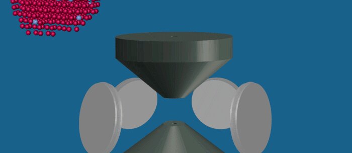
Animation of the creation of X-rays by a sample and the detection of those X-rays by the four detectors in the X-Ant-EM microscope.