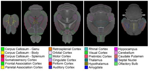
C57BL6 brain atlas
In this study, male WT C57BL/6 J mice and male transgenic APPKM670/671NL/PS1L166P mice were used. Age between 2,4,6 and 8months.
For this atlas, high-resolution 3D anatomical image was acquired using a T2-weighted 3D RARE sequence in horizontal orientation on a 7T Pharmascan (Bruker, Preclinical MRI | Magnetic Resonance Imaging | Research | Bruker).
The following parameters were used: TR = 3185 ms, TE = 44 ms, and spectral width = 50 kHz, averages = 1, RARE factor = 8, matrix size = (265 × 64 × 50), FOV (20.5 × 13.0 × 10.0) mm3, resolution = (0.080 × 0.203 × 0.200) mm3, and total acquisition time = 21 minutes.
A study-based atlas was constructed with Advanced Normalization Tools software (ANT, Home · ANTsX/ANTs Wiki · GitHub) using 25 randomly selected 3D T2-weighted MRI datasets across both genotypes and all ages.
We delineated 23 grey matter and white matter regions of interest (ROIs) on this atlas using AMIRA (ThermoFisher Scientific, Amira Software | Life Science Research | Thermo Fisher Scientific - BE )

The link the the in-house C57BL6 mouse MRI atlas can be found here