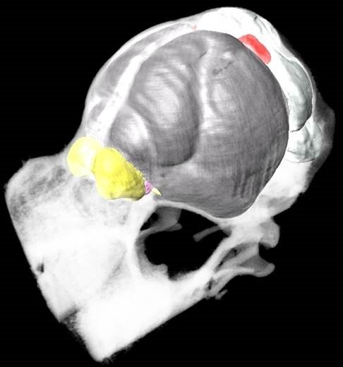
The Mustached Bat brain atlas and its manual can be freely downloaded as zip-file and can be interactively explored with AMIDE, AMIRA 5.4.5, MRIcro, MRIcron, MRIcro-GL, or any other common MRI visualization program.

Substantial knowledge of auditory processing within mammalian nervous systems emerged from neurophysiological studies of the mustached bat (Pteronotus parnellii). Mustached bats retrieve precise information about the velocity and range of their targets through echolocation. This species is also highly social and vocal, emitting a vast array of conspecific communication sounds that are constrained by quasi-syntactical rules. Such high acoustic processing demands were likely the evolutionary pressures driving the hypertrophy of auditory structures at the peripheral (cochlea), metencephalic (cochlear nucleus), mesencephalic (inferior colliculus), diencephalic (medial geniculate body of the thalamus), and telencephalic (auditory cortex) levels in this species. Auditory researchers stand to benefit from a three dimensional brain atlas of this species, due to this considerable contribution to auditory neuroscience. Our MRI-based atlas was generated from several sets of MRI data (9.4 Tesla Bruker scanner) acquired from an ex-vivo male mustached bat’s brain, including a detailed T2-weighted 3D-RARE scan [(59 x 63 x 85) mm3] as well as a fractional anisotropy, radial diffusivity, and track density imaging maps based on super resolution diffusion tensor images [(78) mm3] reconstructed from a set of low resolution diffusion weighted images using Super-Resolution-Reconstruction (SRR). Estimates of auditory cortex location in this species were derived from published neuroanatomical maps superimposed onto a 3D reconstruction of the bat’s cortex. This MRI-based neurological atlas increases the likelihood of performing successful neuroimaging, electrophysiological, and microinjection studies in this species.