Cornea-on-chip
A microscale model with huge potential
Constructing a human cornea on a chip may still sound futuristic, but for doctoral researcher Joris Van Meenen, it is part of his everyday routine. Within his research group ARGOS (Translational Neurosciences), he is developing a cornea-on-chip model designed to mimic the barrier function of the human cornea. “A cornea-on-chip could, for instance, provide us with new insights about the impact of eye blinking”, Joris explains.
“In theory, testing novel pharmaceuticals directly on humans would, of course, yield the best results, but for ethical reasons that isn’t possible. That’s why, in medical research, we continuously look for models that replicate human physiology as accurately as possible”, Joris says. The cornea-on-chip is part of the emerging organ-on-chip (OOC) field, an innovative area of research that is becoming increasingly popular in preclinical testing of drugs and treatments.
From 2D to 4D
An organ-on-chip is a miniature chip containing tissue and embedded with microfluidic channels. “Through these channels, we can simulate fluid flows similar to those in the human body. This allows us to feed the cells or administer drugs to study their effects on the tissue”, Joris explains.
Drug development has undergone a remarkable evolution in recent decades. “Initially, researchers worked with 2D cultures, in which cells were grown on a plastic surface. Later came the 3D cultures, composed of multiple cell types and connective tissue. But even those models remain static, whereas the human body is inherently dynamic”, Joris says.
Today, organ-on-chip technology can be seen as a type of ‘4D culture’. “OOC technology combines the three-dimensional structure of tissue with dynamic properties, enabling us to mimic human physiology more realistically”, he adds.
In this way, OOC technology forms a promising intermediate step between simple in-vitro tests and more complex animal models. “Some labs already connect multiple organ-on-chip systems to each other. Think, for example, of a gut-on-chip that models drug absorption in the intestines, connected to a liver-on-chip that shows how those drugs are metabolised. Step by step, we are moving towards a human-on-chip, a system that approximates the complexity of an animal model as closely as possible”, Joris explains.
Cornea-on-chip
Organ-on-chip technology also holds considerable potential in ophthalmology, particularly because the human cornea is so complex. “You can use animal models for corneal research, but they are not always representative: animal corneas can be thicker, the cells grow differently, or the barrier function varies. That’s why human cells remain the most valuable”, says Joris. A cornea-on-chip could also help reduce painful animal experiments such as the DRAIZE test, a classical test in which substances are applied to a rabbit’s eye to assess irritation.
During his PhD research, Joris focuses on developing a cornea-on-chip that mimics the corneal barrier function, with the aim of measuring how effectively the tissue blocks certain substances. Although such techniques already exist, Joris is introducing an innovative improvement by integrating them directly into the chip. “We assess the barrier function through electrical resistance: the higher the resistance, the stronger the barrier. In traditional setups, the cornea rests on an artificial plastic membrane. But in my research, I found that this membrane can lead to misinterpretation of the data. I solved this by integrating the tissue directly into the chip, without any intermediate membrane”, he explains.
Blinking
A cornea-on-chip may also help explain why we blink. Today, it is still not fully understood how blinking contributes to the healthy functioning of the cornea. “The cornea is composed of different layers, of which the epithelium is the most important barrier. We know this happens because epithelial cells differentiate (Ed. develop into specialised cells), but there is no standard procedure to trigger this process. In a cornea-on-chip, we could mimic blinking by sending fluid flows at specific flow rates through the microfluidic channels. That would allow us to determine whether blinking influences barrier function”, Joris explains.
The same fluid flows can also be automated. “The chips can be connected to pumps that refresh the culture medium or administer new drugs automatically. That automation represents the more translational aspect of my research”, he adds.
Research team
Joris conducts his research under the supervision of Prof. Carina Koppen, Dr. Bert Van den Bogerd, Prof. Sandra Van Vlierberghe en Dr. Debbie Le Blon. In addition, within ARGOS he collaborates with two other doctoral students on the topic of cornea-on-chip. “On the one hand, Banafshe Pishva focuses primarily on biomaterials that can make our corneal model as accurate as possible. On the other hand, Evelyne De Vos examines whether our model truly corresponds to the human cornea, not only in terms of barrier properties, but also to evaluate whether the model could serve as a genuine alternative to the DRAIZE test. This helps us map out what is needed to use the cornea-on-chip model effectively in preclinical testing”, Joris concludes.
---
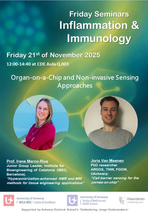
On Friday 21 November, Joris will delve deeper into this topic during the Friday Seminars ‘Inflammation & Immunology’ on the Campus Drie Eiken. In addition, Professor Irene Marco Rius from the Institute for Bioengineering of Catalonia (Barcelona) will give a lecture on her innovative MRI technique, which she applies to small-scale models such as OOC’s.
