16/11/2022 - Charles (ESR #9)

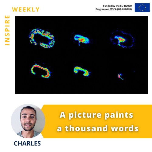
The provided image shows how mass spectrometry imaging can highlight specific metabolites distribution within the tissue (kidney), characterizing its different substructures: cortex (top left), medulla (top right), pelvis (bottom right), and vessels.
Mass spectrometry imaging (MSI) is a powerful technique that enables untargeted investigations into the spatial distribution of molecular species in a variety of samples. It can image thousands of molecules, such as lipids, peptides, proteins, metabolites, and glycans, in a single experiment without any labeling.
Together with my colleague Dustin Krüger ESR#14 from Antwerp University, we started to develop an MSI-related protocol called derivatization, in order to sensitively increase doxorubicin detection within organs like kidney (flyer image), heart, and liver. This study will allow us to evaluate doxorubicin-induced toxicity and will lead to a better understanding of the molecular mechanisms in the organs that metabolize the drug.